


Sm-2010dm Digital Microscope 40X-1000X Digital USB Microscope
Description
Overview
Basic Info.
- Model NO. SM-2010DM
- Magnification 40-1000X
- Function Fogproof
- Usage Digital Microscope
- Structure Refractor Telescope
- Lens Type Prime Lens
- Lens Glass
- Objective Aperture <30mm
- Focusing Type Center Focusing
- Aperture 50-100mm
- Principle Optics
- Mobility Portable
- Number of Cylinder Binoculars
- Shape Single-Lens
- Transport Package Carton Box
- Specification As shown in the figure
- Trademark Shunma / OEM
- Origin Yuyao, Zhejiang, China
- HS Code 9023000000
- Production Capacity 50,000 PCS/Year
Product Description
Product Description
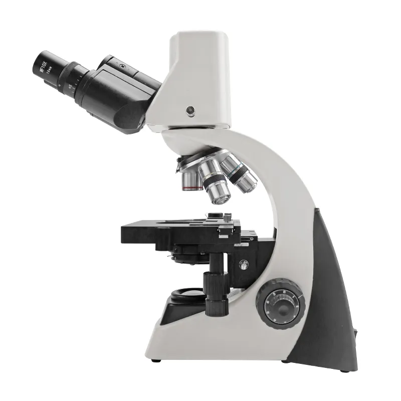
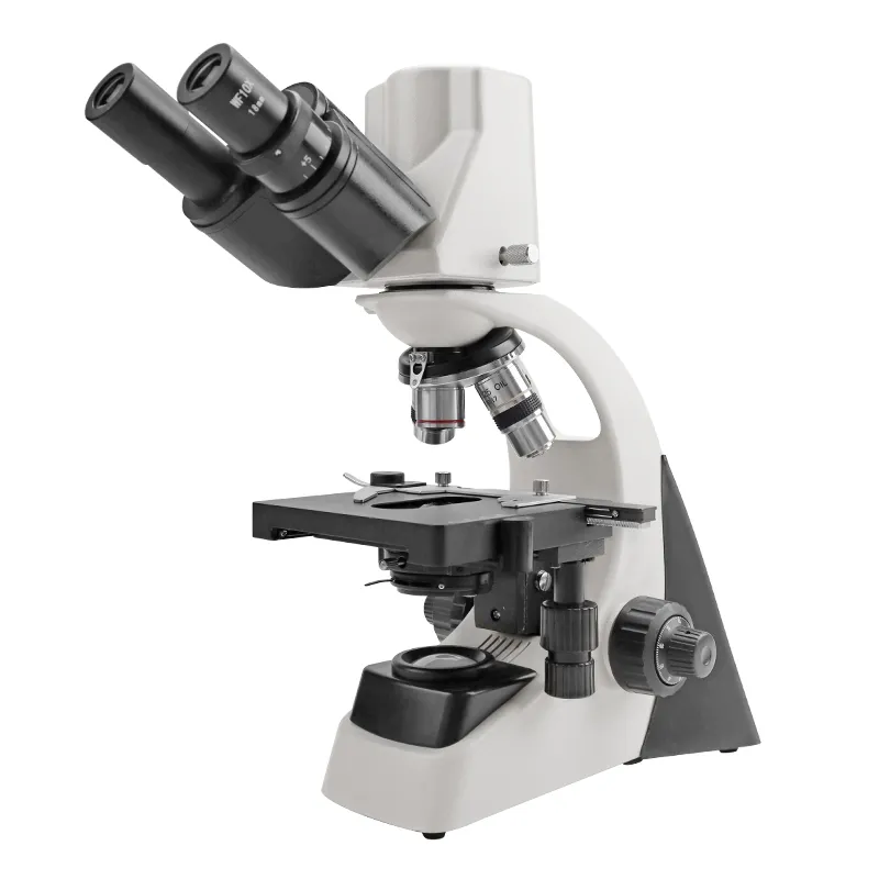
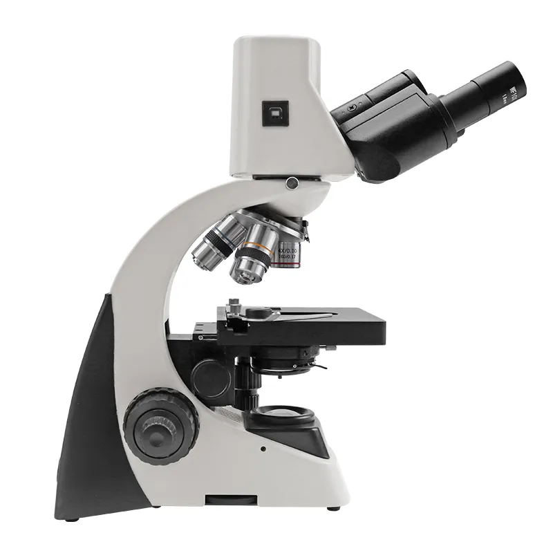
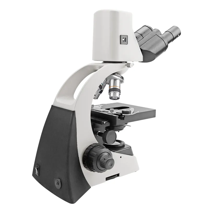
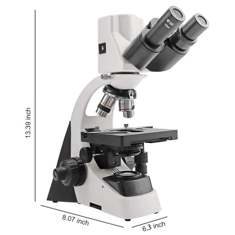
SM-2010DM Digital Microscope (40X-1000X) is a digital USB microscope. It is the successful integration of photoelectric technology and the embodiment of the latest science and technology. Because of its unique image gathering function and strong processing function, it is widely used in schools, hospitals, and research fields. It relieves users from the fatigue of traditional microscopic observation and becomes the best choice for exploring the micro-world.
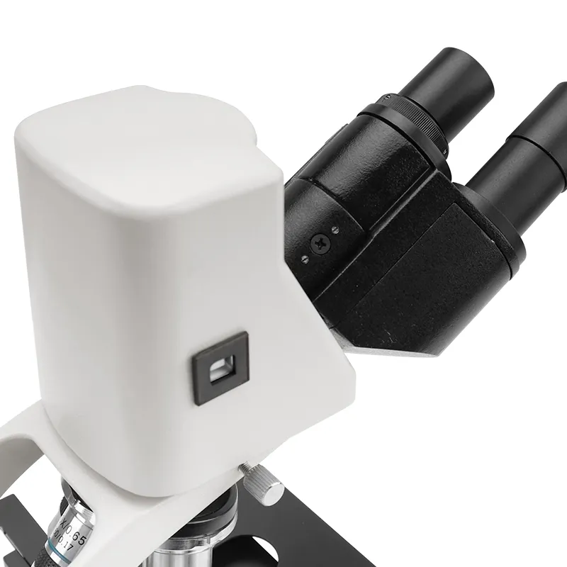
The built-in digital photography system: It is a brand-new micro imagery system that includes a USB2.0 color digital camera with 1.3M pixels. It allows you to obtain any fine information of the micro-image. The image processing system: With the help of a computer, the software can complete the image's previewing, saving, deleting, comparing, measuring, counting, and other functions. The mode of image output: The pure digital signal will be real-time and high-resolution previewed on the computer through USB2.0. Displaying 1280x1024 makes full-screen clear images with high resolution. The pictures will be saved as BMP or JPG files.
Product Specification
| Model | SM-2010DM | 30199004704 | 30199004711 |
|---|---|---|---|
| Total magnification | 40-1000X | 40-400X | 40-1000X |
| Eyepiece | Wide field plane-scope eyepiece WF10X | ||
| Objective | 45mm DIN achromatic objective: 4X, 10X, 40(S), 100(S,Oil) | 45mm DIN achromatic objective: 4X, 10X, 40X(S) | 45mm DIN achromatic objective: 4X, 10X, 40(S), 100(S,Oil) |
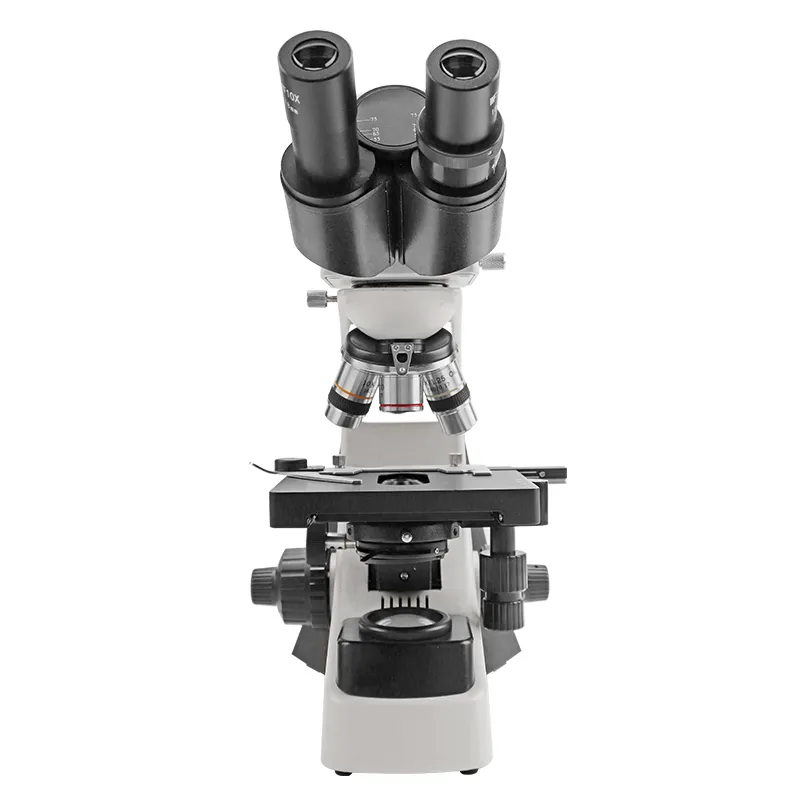
| Eyepiece Tube | Articulated free binocular head, interpupillary distance: 55-75mm; 30° inclined, 360° rotatable | Monocular head, 45° inclined, 360° rotatable | |
| nosepiece | backward quadruple nosepiece | Three-hole converter | Four-hole converter |
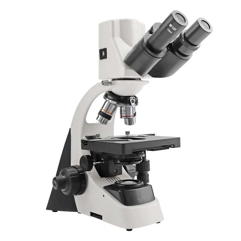
| Illumination | Built-in adjustable brightness halogen lamp 6V/20W (or incandescent, or fluorescent, or LED) | Buit-in incandescent lamp 20W (or halogen, or fluorescent, or LED) | |
| diaphragm | Φ2-Φ30mm iris diaphragm and Φ32mm filter | ||
| condenser | ABBE condenser: N.A. 1.25 | ||
| Adjustable mechanism | Coaxial coarse and fine focusing adjustable mechanism: 14mm | ||
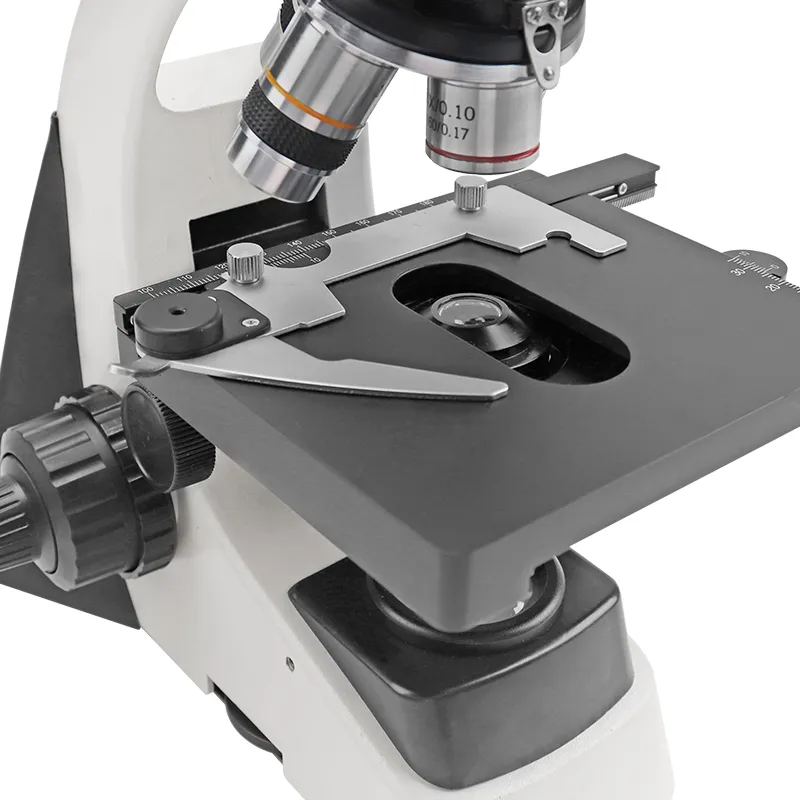
| Working Distance | Double layers mechanical stage: 125x125mm, moving range X-Y: 60x40mm | Double layers mechanical stage: 115*120mm, Moving range X-Y:60*20mm | |
| Other configuration | Built-in digital camera system with a USB2.0 cord and micro-image process software | An external computer and other equipment (with related image processing software) are required, with a photo size of ≥5 million pixels and a video resolution of ≥720 p/30 fps | External projectors, all-in-one machines and other equipment (supporting relevant image processing software) are required. Photo ≥ 14 million pixels, video resolution ≥ 1080 p/30 fps |
SM-2016DM Digital USB Microscope
Objective Lens: 4x, 10x, and 40x (retractable) mounted on a revolving objective turret. LCD Screen: 3.5" LCD screen; displays 5 megapixel images that can be saved to internal memory and to an SD card. Total Magnification (Min.): 40x, 100x, 400x; Stage: includes locked-on stage clips that holds the glass slides; comes with a focus stop to prevent the objective lens from breaking; Includes minimum of 6 position diaphragm; Built-in minimum of 120 MB Memory; With SD card slot. Camera captures 5 megapixel CMOS images or higher; With analog output to connect TV or LCD projector;
Installation
- Requirements for the working environment of the microscope:
- Room temperature: 0 degrees - 40 degrees, maximum relative humidity: 85%
- High temperature: High temperature will cause the microscope to grow mold, mold, and damage the instrument
- Avoid rotating the microscope in a dusty environment, and apply plastic when the microscope is not in use. Cover to cover it.
- The digital USB microscope should be placed in a place without vibration.
- Input voltage check: The input voltage indicated by the adapter should be consistent with the power supply voltage, otherwise it will cause serious damage to the microscope.
- Eyepiece: Insert the eyepiece into the eyepiece brief.
- Objective lens: Lower the stage to the lowest limit, and then screw the objective lenses of various magnifications into the object mirror converter and tighten.
- Condenser: The condenser is already installed when the product leaves the factory.
- Concentrator: The concentrator has been installed and positioned at the time, the concentrator is screw-mounted, and can be rotated counterclockwise out to remove.
- Color filter: When a color filter is required, place the color filter on the variable aperture bracket under the condenser.
Operate
- Insert the eyepiece into the eyepiece, and screw the objective lens into the converter according to the order of different magnifications. When observing, place the specimen in the center of the stage, clamp it with the glass claws, first use the low-power objective to find the image of the observed object, move the observed object to the center of the field of view, and then gradually transfer to the high-power objective. Observation, and at the same time use the micro-hand wheel for fine focusing until the object image is clear. When using the 100X oil immersion objective, it is necessary to fill the space between the front of the objective lens and the specimen plane with fir oil, and there should be no air bubbles in the oil droplets. After the oil lens is used, it should be wiped off with a small amount of xylene immediately to prevent the fir oil from solidifying over time. Not easy to remove.
- In order to obtain a bright and clear object image during observation, attention must be paid to the adjustment of the illumination system. When using objective lenses with different magnifications, the condenser lens should be adjusted and the aperture aperture should be changed to make the numerical aperture suitable for the objective lens. Generally, in other words, when the magnification of the objective lens increases, the condenser must move upward, and the aperture diaphragm is designed for adjusting the numerical aperture, not for adjusting the brightness. Typically, the aperture aperture is opened to 70-80% of the objective's exit pupil until a clear object can be seen in the field of view.
- When a large number of specimens of the same type are to be viewed, the limit screw can be adjusted to the appropriate position. And lock it with a nut, so that after replacing the specimen, directly adjust the coarse handwheel until the first specimen can be seen. To the position of a clear image, it can simplify the operation and reduce the work intensity.
Note: Specimens of the same type refer to sections, coverslips, and slides with the same thickness.
Product Feature and Application
The digital USB microscope can be used in medical and health institutions, universities, secondary schools, technical schools and other units for biological. It is used for bacteriological observation, teaching and professional research, clinical experiment and routine medical examination. This product has a novel shape and adopts a variety of advanced structures and technologies to make the instrument easy to use, safe and reliable.
Maintenance and Precautions
- Be careful when unpacking to prevent the lens and other accessories from falling and being damaged.
- All lenses have been calibrated and adjusted, please do not disassemble them by yourself.
- The objective lens changer and the coarse and fine focus adjustment mechanism have a compact structure, please do not disassemble it easily.
- The instrument should be kept clean, the dust should be removed frequently, and the sliding parts should be regularly coated with a small amount of non-corrosive grease. Special care should be taken not to contaminate the optics when cleaning.
- The instrument should be placed in a cool, dry place, and must be covered with a dust cover after use.
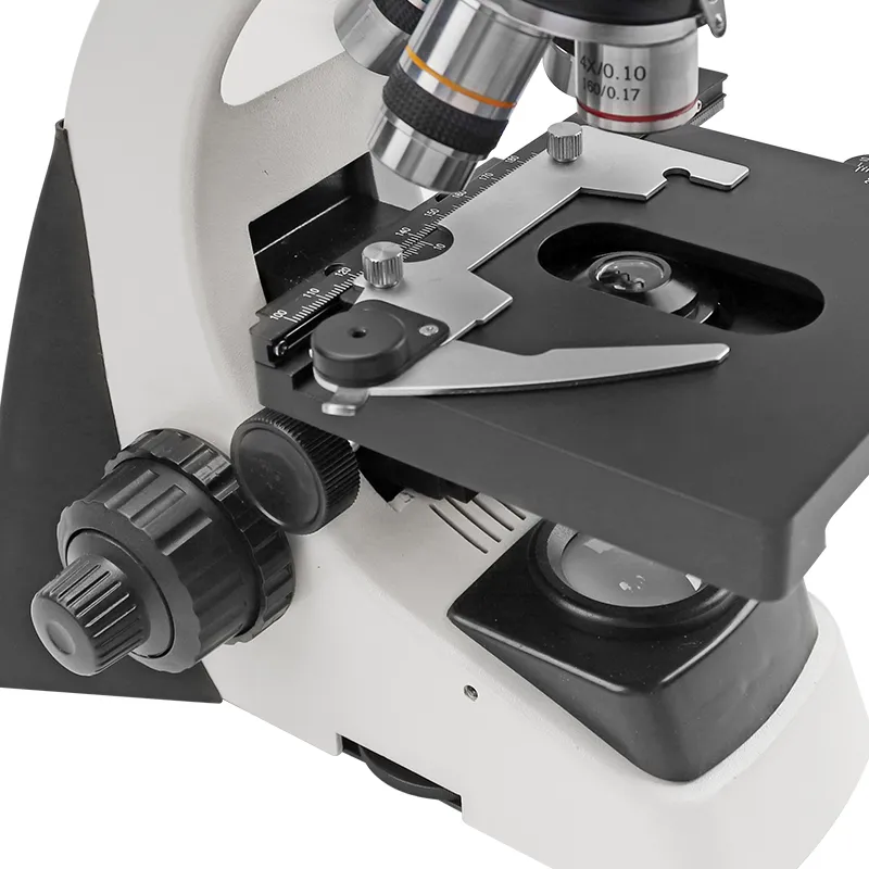
Detailed Photos


You may also like


${currentPro.title}